Thermal Imaging
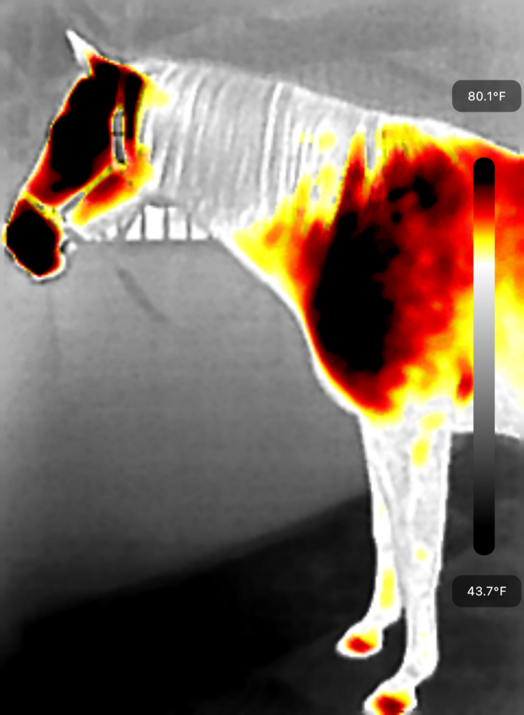
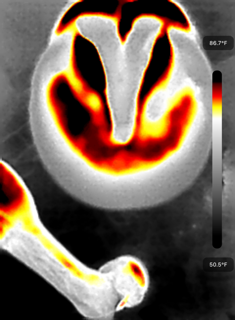
Thermal Imaging, also known as Thermography, is a non invasive technique used to measure the differences in surface temperature.
It can show areas of heat, inflammation, infection and cold or reduced blood flow in the muscular, vascular, skeletal and nervous systems.
Thermal Imaging Cameras are 40 times more sensitive than the human hand (detecting temperature differences of less than 0.05 degrees centigrade) and can detect the location and changes before they can be externally palpated or visually identified!
Ask Chelsea about imaging for your dogs and cats!
For pet owners who are seeking a non invasive and cost effective way to prevent, or to gain more insight before more invasive modalities, Thermal Imaging is a great place to start.
BENEFITS and REASONS TO UTILIZE THERMAL IMAGING:
- Non invasive, objective and supportive! Poses no health risks!
- Post-op monitoring – evaluate the possibility of infection as well as overall recovery of an injury or surgery.
- Pre Purchase Exams – when buying a horse, we want to believe that we are being told the truth about their history. Imaging doesn’t lie!
- Muscle Pain – Injury or tears will always have heat present
- Back Problems or Pain – will show where extra heat and pressure is coming from with the saddle and if this is consistent with where the heat or soreness is without the saddle. ( I also have the impression pad which proves saddle fit)
- Joint and Skeletal Issues – stress fractures and bone chips show up easily on thermal images as increased focalized heat zones. Acute joint injuries or degenerations tend to show up with increased heat initially. Over time, as the problem progresses or becomes chronic, vasoconstriction leads to the area becoming cooler.
- Tendon and Ligament Issues – heat will show in these areas
- Lameness – founder and laminitis have heat associated
- Hoof balance/imbalance, abscesses, bruising and thrush
- Dental Issues
- Hormone Issues or Sheath needing to be cleaned
Preparing for Thermal Imaging
In order to get the best results possible, the following list of requirements must be met
- Avoid direct sunlight or drafty areas
- Fluorescent lighting is preferred or else do not take images directly under incandescent lighting
- Animal must be dry prior to imaging. Sweat or water creates artificial heat zones.
- Do not use any sprays, gels, liniments or give a bath the day of imaging
- Remove all leg wraps, blankets, saddles, restraints, cribbing collars and fly masks at least two hours prior to imaging
- Groom animal at least two hours ahead of time to remove any dirt, mud or manure from body and hooves. Do not do this within two hours of imaging.
- Do not brush, massage, stroke or pet animal while it is being imaged
- Have horse cross tied, if available
- If imaging to assess saddle fit, do not oil the saddle within 48 hours of imaging
- Hooves must be picked out, clean and dry
- Braid tail and place in tail bag if available
- Keep horse out of direct sunlight for at least two hours prior to imaging
- Disclose any drugs animal is on within the last 48 hours and unless advised by the vet for a specific condition, avoid anti inflammatories
- Make note of scars, cuts, bites, rubs etc.
- Reduce any stressors as much as possible as excessive stress may increase thermal activity.
- Images can be taken in a stall, aisle way or open area where they can be taken easily.
What are we looking for?
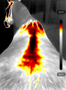
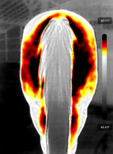
The camera will show the temperature at the hottest and coolest areas on the body. A warmer or hotter area means that there is more blood flow to that region. Heat does not always mean that something is wrong. For example, the nose, mouth and eyes will always show heat. We are looking for symmetry (sameness) on each side of the body. If there are areas that are assyemtrical, this would be cause for a raised eyebrow. Like in this following photo …. we can see that there is something going on, on the right side hamstrings.
I would then be able to target therapy in this area, know that the horse might be reactive here, and monitor this for the future. When the horse is compensating for pain, it will use other body areas and this may create increased thermal activity at a spot that is NOT the original injury site. This does not necessarily mean there is a problem, but should be monitored to avoid further imbalance. Taking a few images after therapy can help show the benefit of the therapy and how the patterns changed. Like in this example of a horse’s neck with a lot of tension and pain that was dispersed and helped with the muscle tape I added.
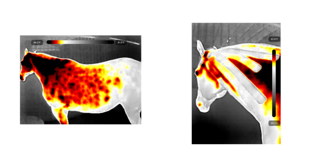
I am not a veterinarian and I do not diagnose or prescribe. The imaging I take is simply for your own knowledge and to take to your veterinarian or equine radiologist for further readings.
How long does imaging take? About 15-20 minutes
How many images will I get? At least 20-25
What do I do with the images? You can keep these images for comparison over time or you can take them directly to your vet if using them in conjunction with other treatments or testing.